
Battlefield Injuries Illustrated by Charles Bell
 L0022552 Watercolour: soldier with left arm missing
Credit: Wellcome Library, London. Wellcome Images
[email protected]
http://wellcomeimages.org
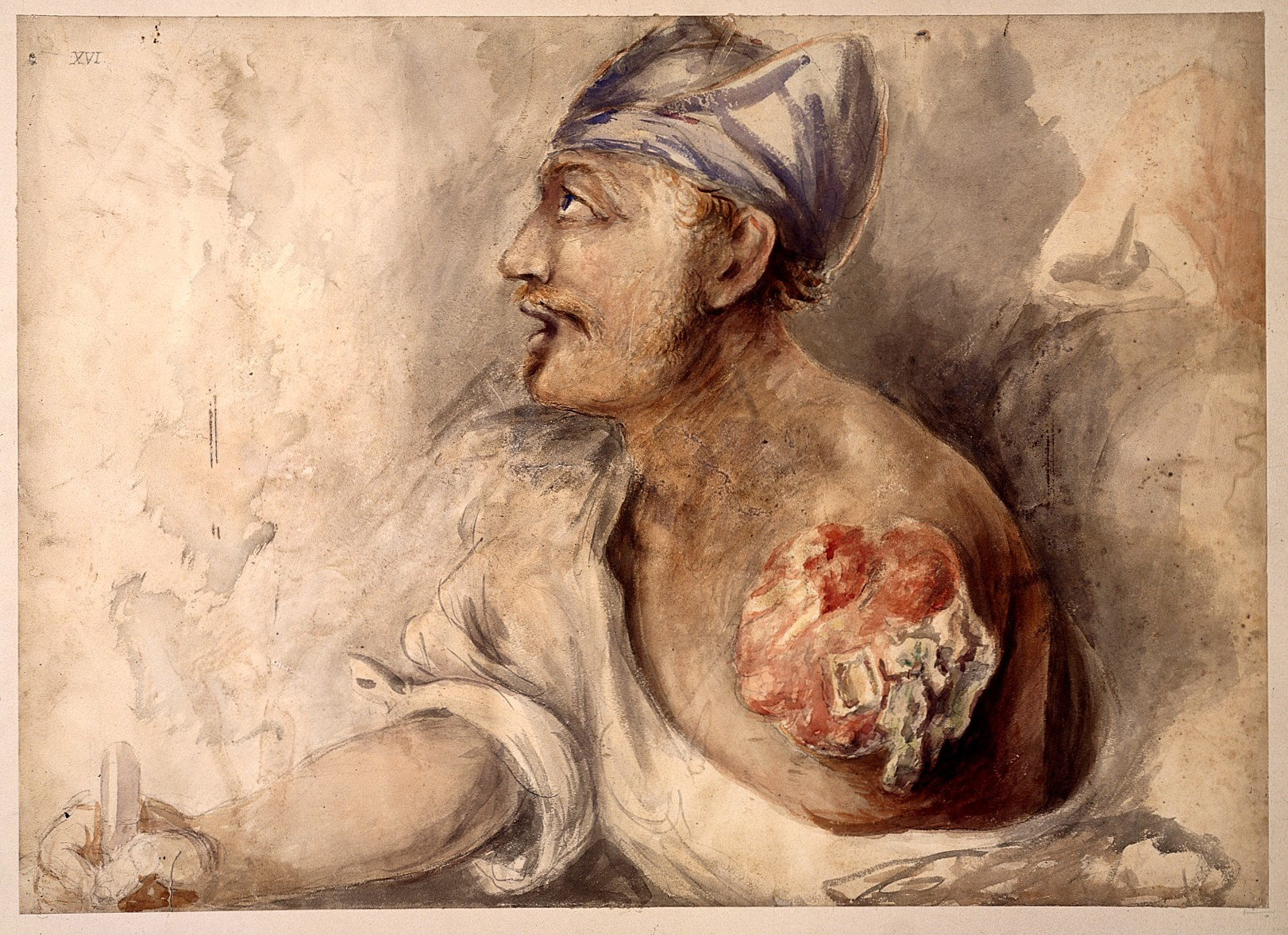
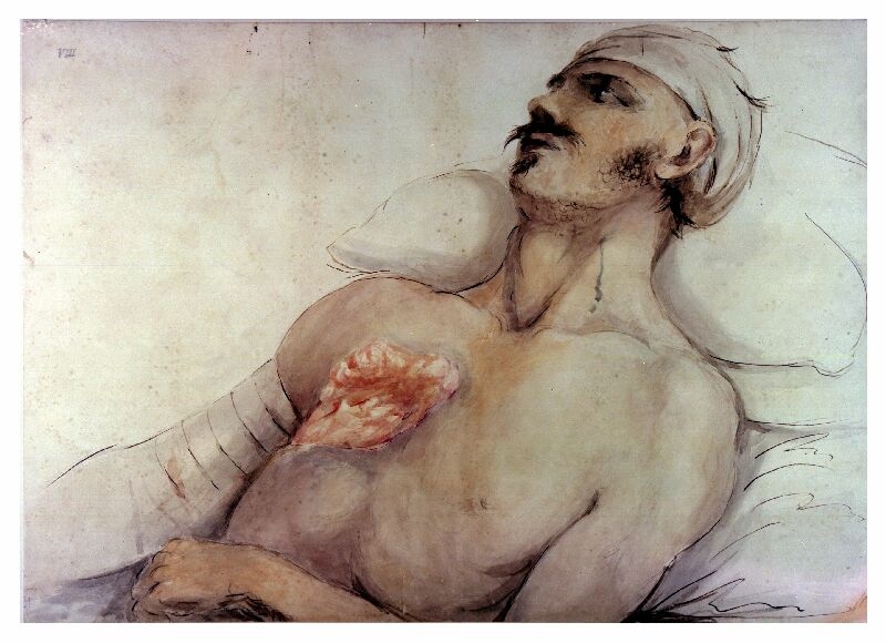
Soldier with left arm missing, bandaged head, with quill in right arm. Wounded at the battle of Waterloo.
Watercolour
1815 By: Charles BellPublished: -
Copyrighted work available under Creative Commons Attribution only licence CC BY 4.0 http://creativecommons.org/licenses/by/4.0/
L0022552 Watercolour: soldier with left arm missing
Credit: Wellcome Library, London. Wellcome Images
[email protected]
http://wellcomeimages.org
Soldier with left arm missing, bandaged head, with quill in right arm. Wounded at the battle of Waterloo.
Watercolour
1815 By: Charles BellPublished: -
Copyrighted work available under Creative Commons Attribution only licence CC BY 4.0 http://creativecommons.org/licenses/by/4.0/
 Charles Bell watercolour after Waterloo.


This entry includes graphic illustrations of war injuries which some may find upsetting.
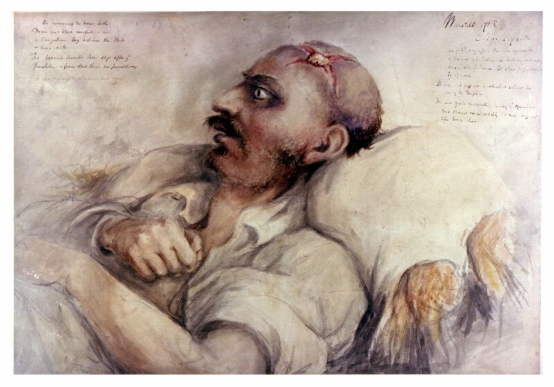
This is a watercolour painting by Charles Bell, a British Army surgeon who helped to treat the wounded after the Battle of Waterloo. His haunting paintings are stark reminders of what early 19th century battle injuries were like. They are rare images executed by a practising surgeon and artist, which was an unusual combination. While his portraits powerfully show the agony and trauma experienced by these young men, they also describe their wounds with anatomical accuracy.
Charles Bell (1774 – 1842) was born and grew up in Edinburgh. While studying medicine at Edinburgh University, he also took drawing classes to develop his artistic skills. In 1804 he moved to London where he would become an accomplished surgeon and anatomist.
10 days after the Battle of Waterloo, in June 1815, Bell joined the military surgeons working tirelessly to treat the thousands of soldiers suffering from terrible wounds inflicted by cannon and musket balls, sabres and other devastating weapons of war. He documented his experiences through sketches, and used these to later paint detailed watercolours. In the absence of photography, Bell’s illustrations and accompanying notes were used for teaching both military and civilian medics.
The main image shows a British soldier called Wanstall, who had suffered a gunshot wound to the skull. The cross wound in Wanstall’s head with the perfect circle in the centre is evidence of trepanning. In this procedure, the surgeons would cut a circle out of the skull to remove pressure on the brain.
The second image illustrates a British soldier whose left arm has been shot away by a cannon ball. In this painting, we can see he is clearly in pain. In his remaining hand he grasps a rope to pull himself up the bed. His terrible wound is still open and would have been difficult to close because of the swelling. The image shows an arterial ligature (tie) applied to the main artery supplying blood to the soldier’s arm to stem the bleeding. This was probably an emergency procedure performed on the field.
The third image shows an unknown soldier with a chest wound.
Did you know..?
Fascinated by the brain and nervous system, Bell was the first to document the difference between motor and sensory nerves in the spinal cord. The relatively common condition of facial paralysis, known as Bell’s palsy, is named after him.
Watch our video about anatomy and the work of Charles Bell.
Use our Classroom resources to investigate this object and the theme of Medicine further.
Highlights:
- Using objects, artworks and other sources to find out about the past
- What factors drove change in medical practice during the Age of Revolution?
And much more…
Related Objects
-
Curatorial info
- Originating Museum: Army Medical Services Museum
- Production Date: 1815-1817
- Creator: Charles Bell
- Technique: Watercolour & charcoal
-
Use this image
You can download a higher resolution image below, but please note the conditions of the licence.
- Rights Holder: Copyright Army Medical Services Museum.
- License Type: All Rights Reserved
Find it here
This object is in the collection of Army Medical Services Museum